Engineered microvessels provide 3-D test bed for human diseases

University of Washington bioengineers have developed the first structure to grow small human blood vessels, creating a 3-D test bed that offers a better way to study disease, test drugs and perhaps someday grow human tissues for transplant.

?In clinical research you just draw a blood sample,? said first author Ying Zheng, a UW research assistant professor of bioengineering. ?But with this, we can really dissect what happens at the interface between the blood and the tissue. We can start to look at how these diseases start to progress and develop efficient therapies.?

Zheng first built the structure out of the body?s most abundant protein, collagen, while working as a postdoctoral researcher at Cornell University. She created tiny channels and injected this honeycomb with human endothelial cells, which line human blood vessels.

During a period of two weeks, the endothelial cells grew throughout the structure and formed tubes through the mold?s rectangular channels, just as they do in the human body.

When brain cells were injected into the surrounding gel, the cells released chemicals that prompted the engineered vessels to sprout new branches, extending the network. A similar system could supply blood to engineered tissue before transplant into the body.

After joining the UW last year, Zheng collaborated with the Puget Sound Blood Center to see how this research platform would work to transport real blood.

Microfluidic vessel networks (credit: Y. Zheng et al./PNAS)

The engineered vessels could transport human blood smoothly, even around corners. And when treated with an inflammatory compound, the vessels developed clots, similar to what real vessels do when they become inflamed.

The system also shows promise as a model for tumor progression. Cancer begins as a hard tumor but secretes chemicals that cause nearby vessels to bulge and then sprout. Eventually tumor cells use these blood vessels to penetrate the bloodstream and colonize new parts of the body.

When the researchers added to their system a signaling protein for vessel growth that?s overabundant in cancer and other diseases, new blood vessels sprouted from the originals. These new vessels were leaky, just as they are in human cancers.

?With this system we can dissect out each component or we can put them together to look at a complex problem. We can isolate the biophysical, biochemical or cellular components. How do endothelial cells respond to blood flow or to different chemicals, how do the endothelial cells interact with their surroundings, and how do these interactions affect the vessels? barrier function? We have a lot of degrees of freedom?,? Zheng said.

The system could also be used to study malaria, which becomes fatal when diseased blood cells stick to the vessel walls and block small openings, cutting off blood supply to the brain, placenta or other vital organs.

?I think this is a tremendous system for studying how blood clots form on vessels walls, how the vessel responds to shear stress and other mechanical and chemical factors, and for studying the many diseases that affect small blood vessels,? said co-author Dr. José López, a professor of biochemistry and hematology at UW Medicine and chief scientific officer at the Puget Sound Blood Center.

Future work will use the system to further explore blood vessel interactions that involve inflammation and clotting. Zheng is also pursuing tissue engineering as a member of the UW?s Center for Cardiovascular Biology and the Institute for Stem Cell and Regenerative Medicine.

Ref.: Ying Zheng et al., In vitro microvessels for the study of angiogenesis and thrombosis, PNAS, May 29, 2012

Source

3D blood vessels could aid artificial organs

Growing artificial organsMovie Camera might help solve the transplantation shortage, but one major hurdle still exists: it is difficult to get blood vessels to grow all the way through a large organ. A gel that allows blood vessels to grow in precise shapes and respond to human cells in a manner similar to natural vessels might help jumpstart that process.

Ying Zheng at the University of Washington in Seattle and colleagues injected human endothelial cells ? which line blood vessels ? into tiny channels within a collagen gel.

The endothelial cells spread throughout the channels, which were only micrometres in width, and formed hollow, three-dimensional tubes, or microvessels. When the researchers pumped blood into the system, it moved through the microvessels without sticking. It could even flow smoothly around 90 degree bends.

The researchers then added a series of proteins involved in inflammation. They found that the proteins caused the blood to clot inside the microvessels, just as it would in the body. Because the system reacted to these stimuli in the same way as a natural vascular system would, Zheng says, it might one day be useful for screening drugs.

When the group injected human brain and muscle cells into the gel, along with proteins that stimulate blood vessel growth, the microvessels showed that they could branch and integrate with the two types of tissue.

Because the channels can be directed into any shape, bioengineer Linda Griffith of Massachusetts Institute of Technology is hopeful that the system can model complex vascular systems such as the blood-brain barrier, which is difficult to study in living animals. Additionally, she adds, researchers could study how cancers metastasise by putting other cell types, such as bone or liver cells, into the channels along with cancerous cells.

Zheng says that the next step is to use the system as a starting point for an artificial organ. Drawing the channels in the right shape will allow the organ to have an adequate blood supply throughout.

Journal reference: Proceedings of the National Academy of Sciences

Source

No more needles: new device images blood flow non-invasively

Israel Institute of Technology (Technion) researchers have developed a device that provides high-resolution images of red and white blood cells in vivo and does an instant diagnosis ? simply by shining a light on the skin.

By eliminating the long wait time for blood test results, the new microscope might help spotlight warning signs, like high white blood cell count, before a patient develops severe medical problems. The portability of the device could also enable doctors in rural areas without easy access to medical labs to screen large populations for common blood disorders.

As a test, the researchers imaged the blood flowing through a vessel in the lower lip of a volunteer. They successfully measured the average diameter of the red and white blood cells and also calculated the percent volume of the different cell types, a key measurement for many medical diagnoses.
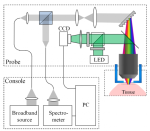
The device relies on a technique called spectrally encoded confocal microscopy (SECM), which creates images by splitting a light beam into a spectrum ? a line from red to violet. To scan blood cells in motion, a compact probe is pressed against the skin of a patient and the rainbow-like line of light is directed across a blood vessel near the surface of the skin.

The blood cells scatter light, which is collected and analyzed. The color of the scattered light carries spatial information, and computer programs interpret the signal to create images of the blood cells at subcellular resolution (.7 micron lateral. 1.5 micron axial).

Currently, other blood-scanning systems with similar resolution exist, but they are far less practical, relying on bulky equipment or potentially harmful fluorescent dyes that must be injected into the bloodstream.

?An important feature of the technique is its reliance on reflected light from the flowing cells to form their images, thus avoiding the use of fluorescent dyes that could be toxic,? says Lior Golan, a graduate student in the Technion biomedical engineering department. ?Since the blood cells are in constant motion, their appearance is distinctively different from the static tissue surrounding them.?

The researchers are working on a second-generation system with higher penetration depth. It might expand the range of possible imaging sites beyond the inside lip, which was selected as a test site since it it?s rich in blood vessels, has no pigment to block light, and doesn?t lose blood in trauma patients.

?Currently, the probe is a bench-top laboratory version about the size of a small shoebox,? says Golan. ?We hope to have a thumb-size prototype within the next year.?

Source

Koç University is accepting MSc (and PhD) degree students to study biomedical engineering and science.

Students who want to improve patient?s well-being and save lives are encouraged to apply as early as possible Committed to be a top BME program of the world, KU-BME is the only biomedical science and engineering program in Turkey, which is run jointly between the Medical School, Science and Engineering, at the same campus !

Guided by the contemporary BME graduate education, you will be supervised by worldrenowned faculty and be trained to be the leader of your research field.

http://gsse.ku.edu.tr

Large-Scale Simulation of Human Blood Is Boon to Personalized Medicine

Having a virtual copy of a patient’s blood in a computer would be a boon to researchers and doctors. They could examine a simulated heart attack caused by blood clotting in a diseased coronary artery and see if a drug like aspirin would be effective in reducing the size of such a clot.

“Blood platelets are like computers in that they integrate many signals and make a complex decision of what to do,” said senior author Scott Diamond, professor of chemical and biomolecular engineering in the School of Engineering and Applied Science. “We were interested to learn if we could make enough measurements in the lab to detect the small differences that make each of us unique. It would be impossible to do this with the cells of the liver, heart or brain. But we can easily obtain a tube of blood from each donor and run tests of platelet calcium release.”

Read More